


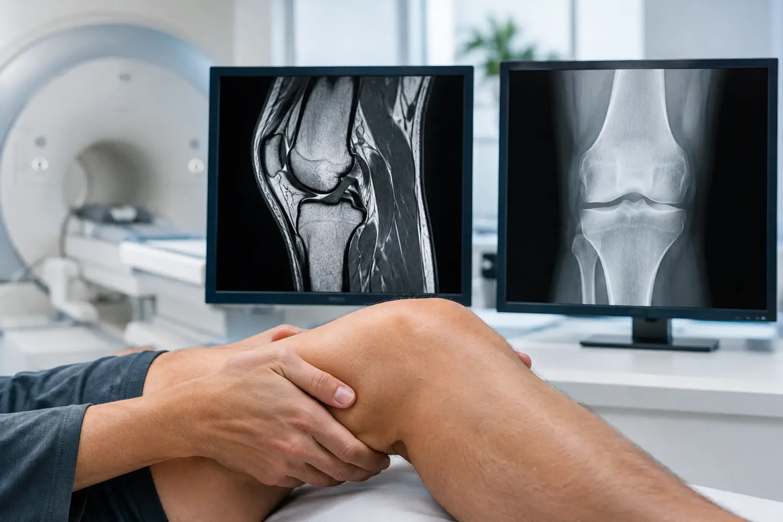
A painful knee can make ordinary decisions feel disproportionately difficult: taking the stairs, getting out of a car, walking the dog or returning to exercise. When imaging is recommended, the question is often whether a knee MRI versus X-ray will provide the answer. They are not competing tests. Each sees different structures, and the right choice depends on your symptoms, examination findings, medical history and the treatment being considered.
Knee MRI versus X-ray: the essential difference
An X-ray is usually the first imaging test for persistent knee pain, particularly in adults with suspected arthritis, a change in alignment or a previous injury. It uses a small amount of radiation to produce clear images of bone. In a few minutes, it can show joint-space narrowing, bone spurs, fractures, changes in joint shape and the position of a previous knee replacement.
An MRI scan uses a powerful magnetic field and radio waves rather than radiation. It creates detailed images of the soft tissues within and around the knee, including the menisci, ligaments, tendons, cartilage, muscles and bone marrow. It is particularly useful when a specialist needs to understand why pain, swelling, catching or instability is occurring despite an X-ray that is normal or does not fully explain the symptoms.
The question is therefore not which scan is “better”. It is which scan is most likely to answer the clinical question at that stage of your care.
What an X-ray can show clearly
For many people over 45 with gradually increasing knee pain, stiffness and reduced walking tolerance, weight-bearing X-rays provide the most useful starting point. Taking images while standing matters because it shows how the joint performs under load. Arthritis may be more apparent when you are upright than when you are lying down.
X-rays can help identify the degree and pattern of osteoarthritis. A specialist will assess the space between the bones, which reflects the remaining cartilage, as well as bone spurs, cysts, deformity and whether arthritis is mainly affecting the inner, outer or kneecap compartment. This detail helps guide discussions about physiotherapy, activity modification, injections, partial knee replacement or total knee replacement.
They are also valuable after trauma. An X-ray can quickly identify or exclude many fractures and dislocations. For patients who have had joint replacement surgery, serial X-rays are central to monitoring the position of the implants, fixation to bone and signs that may suggest loosening or wear.
However, an X-ray cannot directly show a torn meniscus, cruciate ligament injury or irritation within the bone marrow. It may look relatively reassuring even when a soft-tissue problem is causing substantial symptoms.
When an MRI scan adds value
An MRI is often considered when symptoms suggest an injury to a structure that cannot be seen on X-ray. This may include a twisting injury followed by swelling, a knee that gives way, repeated locking, pain along the joint line, or difficulty returning to sport or work after an injury.
It can show whether the meniscus is torn and, crucially, the type and location of the tear. That matters because not every meniscal tear requires arthroscopy. Some tears are associated with age-related wear and coexist with arthritis; treating the scan result alone is unlikely to relieve symptoms. Others, particularly an unstable tear causing true mechanical locking, may require more urgent specialist assessment.
MRI is also the preferred test for suspected anterior cruciate ligament injury, collateral ligament damage, tendon injury, cartilage defects and certain stress fractures that may not be visible on an early X-ray. It can detect bone bruising and inflammation, which may help explain pain after a significant impact.
For someone with established arthritis, MRI is not routinely necessary before knee replacement. X-rays, a careful examination and a conversation about pain, function and treatment goals are commonly enough to plan care. MRI may be useful when the symptoms and X-ray findings do not match, when there is concern about a different diagnosis, or when a more complex problem needs clarification.
Why the examination still matters
A scan should support clinical judgement, not replace it. Knee imaging frequently identifies changes that may not be the source of pain. For example, degenerative meniscal changes are common in middle age and later life, including in people who have no knee symptoms. Equally, an X-ray may demonstrate arthritis, but the location and character of your pain may point to another contributing problem.
A specialist assessment considers how the pain began, where it is felt, what movements provoke it and how it affects sleep, work, exercise and everyday independence. Examination assesses swelling, range of movement, joint stability, alignment, tenderness and the way you walk. Imaging then helps confirm the suspected diagnosis and decide whether non-operative treatment, an injection, rehabilitation or surgery is appropriate.
This approach avoids two unhelpful extremes: dismissing persistent symptoms because an initial scan is normal, or recommending treatment for an incidental finding that does not fit the clinical picture.
Practical differences between the tests
X-rays are fast, widely available and generally straightforward. You may be asked to stand in different positions so that the images accurately assess joint alignment and compartment-specific arthritis. The radiation dose from a knee X-ray is low, but it should still be used only when clinically justified.
An MRI takes longer, often around 20 to 45 minutes depending on the protocol, and requires you to lie still in the scanner. It does not involve ionising radiation. Some people find the enclosed space uncomfortable, although support is available and many scanners are designed to feel less restrictive than older machines.
You must tell the imaging team about any implanted medical devices, metal fragments, clips or previous surgery. Many modern joint replacements and orthopaedic implants are MRI-compatible, but the scanner can produce image distortion around metal. Specialist MRI techniques can sometimes reduce this artefact when investigating pain after knee replacement, though X-rays remain the usual first-line assessment.
MRI scans are typically more expensive than X-rays and are not always the quickest route to a useful answer. Ordering an MRI before an appropriate examination or basic X-rays can lead to delays, uncertainty and findings that do not change treatment.
Choosing the right test for common situations
If knee pain has developed gradually, is worse with walking or stairs and is accompanied by stiffness, a weight-bearing X-ray is often the logical first investigation. It can establish whether arthritis is present and how advanced it is.
If your knee twisted during sport, swelled rapidly, feels unstable or locks so that it cannot fully straighten, an X-ray may still be needed first to exclude a fracture, but MRI is more likely to clarify ligament, meniscal or cartilage injury. Urgent assessment is sensible after a significant injury, a visibly deformed knee, inability to bear weight, fever with a hot swollen joint, or a calf that is suddenly swollen and painful.
If you have ongoing symptoms after previous knee surgery or replacement, the choice is more individual. X-rays may reveal changes to implant position or bone. Blood tests, aspiration of joint fluid, CT scanning, MRI with metal-reduction sequences or other investigations may be considered depending on the concern. Complex cases benefit from a clear, stepwise plan rather than assuming one scan will provide every answer.
Imaging should lead to a treatment decision
The value of an X-ray or MRI lies in what happens next. For some people, the result confirms that a period of targeted physiotherapy, strength work, weight management or an injection is the most appropriate route. For others, it helps establish whether arthroscopy, ligament reconstruction, partial knee replacement, total knee replacement or revision surgery may offer a realistic improvement in pain and mobility.
At a specialist consultation, you should leave understanding what the images show, what they do not show and how the findings relate to your own symptoms. You should also be told honestly when a scan is unlikely to alter treatment. That clarity matters, particularly when you are weighing a significant procedure against the wish to remain active and independent.
If knee pain is limiting the parts of life that matter to you, the most useful next step is not simply requesting the most detailed scan. It is obtaining a careful assessment that selects the right investigation and turns the result into a considered plan for getting you moving with greater confidence.